Monday, August 23, 2021

Non-Membranous Organelles-Cytoskeleton (Microtubules)

Microtubules


Definition: >>-Non branching & rigid hollow fine tubes >>-Formed of tubulin protein. L.M: Not seen, Needs special stains

 E.M: -Fine tubules: >>-Diameter: 20-25nm >>-Length: Changed by adding or removal of tubulin molecules at their end

Forms of microtubules: >>1-Dynamic: Continuous assembly & disassembly → Rapid reshaping of the cell >>2-Stable: Solid microtubules forming the wall of centrioles & flagella

Functions: >>1. Maintain cell shape e.g. platelets >>2. Elongation & movement of the cell 3. Intracellular vesicular transport >>4. Formation of mitotic spindle during cell division 5. Formation of centrioles, cilia & flagella

Sunday, August 22, 2021

Non-Membranous Organelles (Ribosomes)

Ribosomes


Def: >>-Non-membranous rounded or oval bodies [12 X 25 nm] >>-Formed of strands of rRNA + associated proteins >>-Formed in the nucleolus, then pass to cytoplasm through nuclear pores -Found in large numbers in protein synthesizing cells 

L.M: -Very minute structures (15-20 nm) -Difficult to be seen by 

L.M. -Basophilic when stained by H+E [due to RNA content] 

E.M: -Small electron-dense granules >>-Formed of 2 subunits (Large & Small) Forms: 1-Free: -Single or -In groups connected by mRNA strand → forming spiral chains (polysomes) >>2-Attached: To the outer surface of rER by the large subunit at receptors called [Ribophorins]

Function: Protein synthesis >>1-Free ribosomes: >>-Form proteins for local use in cytoplasm -Common in immature cells [Embryonic & Stem cells] >>2-Attached ribosomes: >>-Form proteins for export [secreted by the cells as enzymes & hormones]


Proteasomes 


 Def: >>-Very small non-membranous cylindrical protein complexes >>-Contain proteases Function: Degrade denatured or nonfunctional proteins

 Considered as a quality control unit to ensure quality of exported proteins

Peroxisomes

 Peroxisomes


 Small membranous spherical organelles

Contain enzymes synthesized in free ribosomes: >>1-Oxidases → Oxidize fatty acids producing H2O2 >>2-Peroxidases as Catalase → Break down H2O2 [Which is damaging to the cell] >>3-Other peroxidases → Inactivation of toxic molecules as some drugs, [as in liver and kidney cells] → Formation of bile acids and cholesterol


Saturday, August 21, 2021

Lysosomes

 Lysosomes


Def: >>-The digestive system of the cell >>-Formed at mature surface of G.A >>-Contain about 40 different hydrolytic enz. [phospholipases, proteases, nucleases,….] >>-Numerous in phagocytic cells [WBCs] 

L.M: -Not stained by H & E -Stained by special histochemical stain [acid phosphates] for enzymes inside 

E.M: -Electron-dense appearance -2 Types are distinguished

1ry lysosomes: -Newly formed -Small homogenous vesicles

2ry lysosomes: -Fusion of 1ry Lysosomes + intracellular macromolecules >>-Heterogeneous vesicles

Types of Secondary lysosomes: >>1-Heterolysosomes: Fusion of 1ry lysosome + Phagosome containing solid food or bacteria >>2-Multivesicular bodies: Fusion of 1ry lysosome + Pinocytic vesicle containing fluid droplets >>3-Autolysosomes: Fusion of 1ry lysosome + autophagic vacuole containing destroyed old organelles [as dead mitochondria, rER, …..], the process is called Autophagy >>4-Residual bodies: 2ry lysosomes containing indigestible remnants

Fate of Residual Bodies: >>1-Exocytosis of their content [Cytostool] >>2-Converted to lipofuscin pigment in long-lived cells [Heart muscles, neurones,……]

Important Notes Cytosolic components are protected from lysosomal enzymes by: >>1-The membrane surrounding lysosomes >>2-The pH of cytosol (~7.2) which is not optimal for enzymes activity

 The optimum pH for enz. activity is (~5.0). So, leakage of lysosomal enzymes isn’t harmful to the cell

Functions of Lysosomes n Digestion of: >>1-Nutrients in cytoplasm >>2-Solid particle (phagocytic vesicles): bacteria & viruses → Immune function >>3-Fluid droplets (Pinocytic vesicles) >>4-Old organelles >>5-Dead cells (autolysis after death)


Golgi Apparatus

 Golgi Apparatus


Def: -The secretory system of the cell [Named after histologist Camillo Golgi who discovered it in 1898]

Well developed in secretory cells

L.M: -Not stained by H&E due to high lipid content in its membranes ® Gives pale area in cytoplasm called “Golgi ghost” or “–ve Golgi image” also

E.M: >>-Many flat membranous saccules, Stacked over each other >>-All saccules contain enzymes and proteins being processed >>-Located near the nucleus >>-Each saccule has two faces

 Immature (Cis) face: Receives transport vesicles from rER

Mature (Trans) face: The shipping area that gives two types of vesicles

Types of vesicles: >>1-Secretory vesicles for exocytosis >>2-Lysosomes 

N.B: Secretory granules: >>-Granules containing concentrated secretory products [up to 200 times than those in rER] until released by exocytosis >>-Zymogen granules: Secretory granules in pancreatic cells with dense contents of digestive enzymes

 Functions of Golgi Apparatus: Completes the function of rER >>1-Concentration and packaging of proteins formed by rER >>2-Chemical modification of proteins >>3-Discharge of secretory vesicles ® Secretions >>4-Formation of lysosomes & isolation of their enzymes >>5-Maintenance of cell membrane & surface coat


Friday, August 20, 2021

Endoplasmic Reticulum

 Endoplasmic Reticulum


Definition: System of intercommunicating channels [Cisternae]  Forming reticulum [Network] in cytoplasm

Rough Endoplasmic Reticulum >> Site Protein forming cells: Plasma cells, Osteoblasts & Pancreas

-:Functions

1Synthesis of protein by ribosomes >>2-Transfer of protein to Golgi apparatus through transfer vesicles >>3-Formation of enzymes of lysosomes >>4-Intracellular pathway

L.M Basophilic cytoplasm [due to rRNA]

E.M Reticulum of parallel intercommunicating cisternae with attached ribosomes  Rough outer surface

Smooth Endoplasmic Reticulum >> Site Lipid forming cells: Liver Endocrine glands: Suprarenal cortex

L.M If abundant >> acidophilic cytoplasm [Normally not seen by L.M] 

E.M Reticulum of anastomosing cisternae without ribosomes >> Smooth outer surface

 Functions -Synthesis of Lipid >>2-Synthesis of steroid hormones >>3-Storage of glycogen [in liver & muscles] >>4-Helps muscle contraction by Ca pump >>5-Detoxification of drugs [in liver]


Mitochondria

 Mitochondria


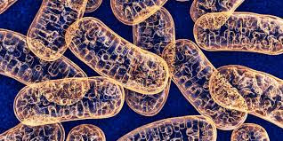
Definition: Power house of the cell [Responsible for cell respiration & energy production] Number: Numerous in active cells >> -Liver cells [contain up to 2000/cell] >>-Cardiac muscle >>-Kidney tubules 

L.M: >>-Granules, rods or filaments. >>-Need special stains: Stains black with iron hematoxylin, stains green with Janus 

E.M: >>-Round or oval vesicles >>-Covered with double membranes separated by inter >>-membranous space >>-Outer membrane is smooth >>-Inner membrane form folds [called cristae] >>-Matrix fills the internal cavity, contains: Lipid, Protein, Carbohydrate, Ca, Mg, DNA, RNA & Oxidative enzymes

Functions: >>1-Cell respiration [Power House]: Contain oxidative enzymes  produce energy [ATP] through Krebs cycle.  50% of energy is stored as ATP  50% is dissipated as heat to keep normal body temperature >>2-Form their own proteins & can divide, as they contain DNA & RNA. 

Role of Mitochondria in Apoptosis [Cell suicide]  On exposure of the living cell to stress [Hypoxia, heat, viruses, DNA damage,…..], Mitochondria release Cytochrome C protein from inner membrane  Activation of proteases enzymes in cytoplasm  Apoptosis


A-Membranous Organelles

Cell Membrane


Definition: Very thin membrane surrounding the cell 

L.M: -Invisible as it is very thin [8-10 nm] -But can be stained by PAS or Ag 

E.M: Trilamellar membrane [Appears as 2 dark parallel lines, separated by light one]

 Molecular structure of cell membrane: >> 1-Lipid Component [30%]

a-Phospholipid molecules

Arranged in 2 layers (bilipid layer)

Each molecule has a head & tail

b-Cholesterol molecules

-Protein Component [60%]

Integral protein [Channel or carrier protein]

Peripheral protein

Carbohydrate Component [10%]: -They are Polysaccharides. -Either attached to protein >> Glycoprotein or to lipid >> Glycolipid -Glycolipid + Glycoprotein >> Cell coat or [Glycocalyx]

Functions of Cell Membrane >> 1-Passive diffusion (for gases, water & ions) according to concentration gradient >> 2-Facilitated diffusion (with carrier): for glucose and amino acids. >> 3-Active transport (needs energy): e.g. Na+-K + pump. >> 4-Selective permeability (by receptors in cell coat): Allows entry of certain substances >>  5-Phagocytosis: Engulfment of solid materials >> 6-Pinocytosis: Engulfment of liquid materials >> 7-Exocytosis: Extrusion of substances outside the cell >> 8-Functions of cell coat: a-Mechanical protection of the cell b-Cell immunity & allergy

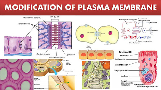
Modifications in Cell Membrane


 Formation of microvilli → Increased surface area e.g: Absorptive cells in small intestine >> Formation of cilia → Move particles above cell membrane e.g: Removal of dust from inspired air in respiratory passages >> Formation of flagella → Helps movement e.g: Tails of sperms

Cytology The Cell


Definition: -The structural & functional unit of all living tissues [Cells have different shapes & sizes]
The Cell is made of: I-Cytoplasm II-Nucleus
I-Cytoplasm 
Formed of
1Cytoplasmic Matrix: Colloidal solution contains proteins, carbohydrates, lipids, minerals & enzymes
2Cytoplasmic inclusions: -Non-living temporary structures -Not essential for the cell vitality. -They are stored substances in some cells, e.g. glycogen, fat & pigments
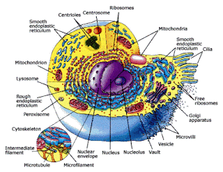
3Cytoplasmic organelles: -Living permanent minute structures -Essential for vital processes: respiration, secretion, digestion
Types
A-Membranous

1Cell membrane
2Mitochondria 
3Endoplasmic reticulum (rough & smooth)
4Golgi apparatu
5Lysosomes

B-Non-membranous

1Ribosomes 
2Cytoskeleton 
 a-Microtubules (centrioles & cilia) 
 b-Filaments (Thin, intermediate & thick) 

Staining

Staining


Tissues & cells are colorless. So, must be stained for examination >> Stains are
 either acidic or basic >> The classic stain used is Hematoxylin + Eosin [H & E] >> Hematoxylin is a basic stain [Stains acidic components like DNA of nucleus] [The affinity of acidic structures to basic stains is called basophilia] >> Eosin is an acidic stain [Stains basic components like proteins, mitochondria, collagen,…] [The affinity of basic structures to acidic stains is called acidophilia]

Some Special Stains for L.M
Vital stain: Staining a living structure inside a living animal 
 Ex.: Staining of phagocytic cells using trypan blue or India ink >> Supravital stain: Staining a living cell outside a living person
 Ex.: Brilliant cresyl blue → Stains reticulocytes (immature RBCs) in a blood film. >> Metachromatic stain: The stain gives a different color from the original one due to chemical combination between the stain & certain structures within the cell [Metachromasia]
 Ex.: Toluidine blue stains granules within mast cell with a violet color. >> Histochemical stains: Using enzyme reaction to demonstrate an enzyme or chemical components of the cell
 Ex.: -Alcian blue: used to detect mucin protein in mucous secreting cells -Acid phosphatase enzymes: used to detect enzymes of lysosomes -Periodic Acid-Schiff reaction [PAS]: used to detect polysaccharides in cell membranes >> Immunohistochemical stain: Based on specific reactions between antigen + antibodies labeled with fluorescent dye to emit visible light [Immunofluorescent technique]


Thursday, August 19, 2021

Microtechniques

Def: Preparation of histologic sections from tissues for microscopic examination >> The ideal micro-technique that preserves structural relations between cells as in living tissue


Paraffin Technique for L.M >> Fresh small tissue samples [1cm] are obtained >> Steps: 1-Fixation: in formalin → Preserve tissue structure 2-Dehydration: in ascending grades of alcohol [50%, 70%, 90% then in 100%] → Removes all water. 3-Clearing: Alcohol is removed in chemical solvents [xylol] → Tissue becomes translucent 4-Infiltration: Tissue is placed in melted paraffin until it becomes completely infiltrated 5-Embedding: Paraffin-infiltrated tissue is placed in a small container with melted paraffin and allowed to harden. 6-Trimming: Paraffin block is trimmed to expose the tissue for sectioning on a microtome. 7-Sectioning: The trimmed hard paraffin block is fixed on a holder and cut into thin sections [3-10 µm] by sharp metal knife in a device called [Microtome]

Advantages of Paraffin Technique >> Rapid technique >> Gives serial sections for research >> Gives thin section easily stained 

Disadvantages: >> Xylol dissolves fat content, so cannot be demonstrated >> The heat destroys enzymes of the cell, so cannot demonstrate chemical components

 N.B: Microtechniques for preparation of a tissue sample for electron microscopic examination differ in some points: 1. Tissue samples are smaller 2. Fixation: in Formalin + Glutaraldehyde 3. Embedding: in Epoxy resin to give harder tissue 4. Sectioning: Very thin sections < 1 µm by glass or diamond knife


Types and Parts of microscopes (Light&Electron)

. Types of microscopy: 1-Light microscopy 2-Electron microscopy


-:Parts of light microscope

Ocular lens [2 Eye pieces]: Magnify the image X 10 

Objective lenses: Magnifies the image X 4, X 10, X 40 & X 100 for higher magnification of more detailed areas

The condenser: Collects and focuses a cone of light that illuminates the tissue slide on the stage

Stage control: Moving the slide

How to calculate the total magnification of light microscopy? >> Total Magnification power of light microscope = >> Magnification power ocular lens [10] X Magnification power of objective lens [4, 10, 40 & 100]

Some important measurements & Units >> Centimeter (cm) = 10 Millimeter (mm) >> Millimeter (mm) = 1000 Micrometer (µm) >> Micrometer (µm) = 1000 Nanometer (nm)

The difference between magnification power & resolution power

 Magnification power = Degree of enlargement >> The maximal magnification power of: -L.M is 1000 times & -E.M is 120,000 times 

Resolution power = The least distance between two points that can be seen as two [not one point] The maximal resolution power of: -L.M is 0.2 µm & E.M is 3 nm

Types of Electron Microscope 1- Transmission Electron Microscopy 2- Scanning Electron Microscopy

Transmission Electron Microscopy >> Based on the interaction of tissue components with beams of electrons passing through the tissue sections >> Produce an image with black, white and intermediate gray regions >> Electron-lucent = White (Bright) areas through which electrons passed >> Electron-dense = Dark areas where electrons were absorbed or deflected

Scanning Electron Microscopy >> Provides a high-resolution view of the surfaces of cells, tissues, and organs. >> The beam of electrons does not pass through the specimen >> The surface of the specimen is spray-coated with a very thin layer of heavy metal (often gold) → Reflects electrons in a beam scanning the specimen. >> The reflected electrons are captured → 3D-Black & white